


 癌症的基本特征包括细胞增殖、血管生成、迁移、凋亡逃避机制和细胞永生等。找到癌症发生过程中这些通路的关键标记物和对应的抗体用于检测至关重要。
 为您推荐一个泛素化位点预测神器——泛素化分析工具,可以为您的蛋白的泛素化位点作出预测和评分。
 细胞自噬受体图形绘图工具为你的蛋白的细胞受体结合位点作出预测和评分,识别结合到自噬通路中的蛋白是非常重要的,便于让我们理解自噬在正常生理、病理过程中的作用,如发育、细胞分化、神经退化性疾病、压力条件下、感染和癌症。
MAP2K3 Antibody
Purified Mouse Monoclonal Antibody
- 产品详情
- 实验流程
Application
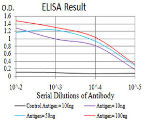
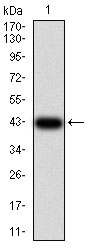
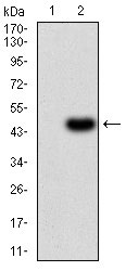
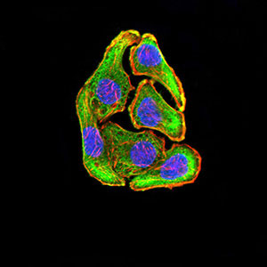
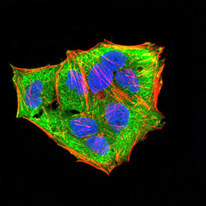
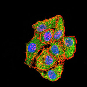
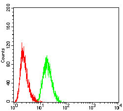
| WB, IHC, FC, ICC, E |
|---|---|
| Primary Accession | P46734 |
| Reactivity | Human |
| Host | Mouse |
| Clonality | Monoclonal |
| Clone Names | 2E12D11 |
| Isotype | IgG1 |
| Calculated MW | 39318 Da |
| Description | The protein encoded by this gene is a dual specificity protein kinase that belongs to the MAP kinase kinase family. This kinase is activated by mitogenic and environmental stress, and participates in the MAP kinase-mediated signaling cascade. It phosphorylates and thus activates MAPK14/p38-MAPK. This kinase can be activated by insulin, and is necessary for the expression of glucose transporter. Expression of RAS oncogene is found to result in the accumulation of the active form of this kinase, which thus leads to the constitutive activation of MAPK14, and confers oncogenic transformation of primary cells. The inhibition of this kinase is involved in the pathogenesis of Yersina pseudotuberculosis. Multiple alternatively spliced transcript variants that encode distinct isoforms have been reported for this gene. |
| Immunogen | Purified recombinant fragment of human MAP2K3 (AA: 1-138) expressed in E. Coli. |
| Formulation | Purified antibody in PBS with 0.05% sodium azide |
| Gene ID | 5606 |
|---|---|
| Other Names | Dual specificity mitogen-activated protein kinase kinase 3, MAP kinase kinase 3, MAPKK 3, 2.7.12.2, MAPK/ERK kinase 3, MEK 3, Stress-activated protein kinase kinase 2, SAPK kinase 2, SAPKK-2, SAPKK2, MAP2K3, MEK3, MKK3, PRKMK3, SKK2 |
| Dilution | WB~~1/500 - 1/2000 IHC~~1:100~500 FC~~1/200 - 1/400 ICC~~N/A E~~1/10000 |
| Storage | Maintain refrigerated at 2-8°C for up to 6 months. For long term storage store at -20°C in small aliquots to prevent freeze-thaw cycles. |
| Precautions | MAP2K3 Antibody is for research use only and not for use in diagnostic or therapeutic procedures. |
| Name | MAP2K3 |
|---|---|
| Synonyms | MEK3, MKK3, PRKMK3, SKK2 |
| Function | Dual specificity kinase. Is activated by cytokines and environmental stress in vivo. Catalyzes the concomitant phosphorylation of a threonine and a tyrosine residue in the MAP kinase p38. Part of a signaling cascade that begins with the activation of the adrenergic receptor ADRA1B and leads to the activation of MAPK14. |
| Tissue Location | Abundant expression is seen in the skeletal muscle. It is also widely expressed in other tissues |
Research Areas
For Research Use Only. Not For Use In Diagnostic Procedures.
Application Protocols
Provided below are standard protocols that you may find useful for product applications.
REFERENCES
1.Hum Mol Genet. 2013 Nov 1;22(21):4438-49.2.Proteomics Clin Appl. 2010 Nov;4(10-11):816-28.
终于等到您。ABCEPTA(百远生物)抗体产品。
点击下方“我要评价 ”按钮提交您的反馈信息,您的反馈和评价是我们最宝贵的财富之一,
我们将在1-3个工作日内处理您的反馈信息。
如有疑问,联系:0512-88856768 tech-china@abcepta.com.
¥ 1,500.00
Cat# AO2234a


