


 癌症的基本特征包括细胞增殖、血管生成、迁移、凋亡逃避机制和细胞永生等。找到癌症发生过程中这些通路的关键标记物和对应的抗体用于检测至关重要。
 为您推荐一个泛素化位点预测神器——泛素化分析工具,可以为您的蛋白的泛素化位点作出预测和评分。
 细胞自噬受体图形绘图工具为你的蛋白的细胞受体结合位点作出预测和评分,识别结合到自噬通路中的蛋白是非常重要的,便于让我们理解自噬在正常生理、病理过程中的作用,如发育、细胞分化、神经退化性疾病、压力条件下、感染和癌症。
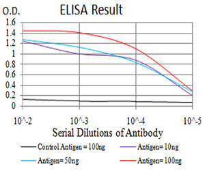
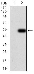
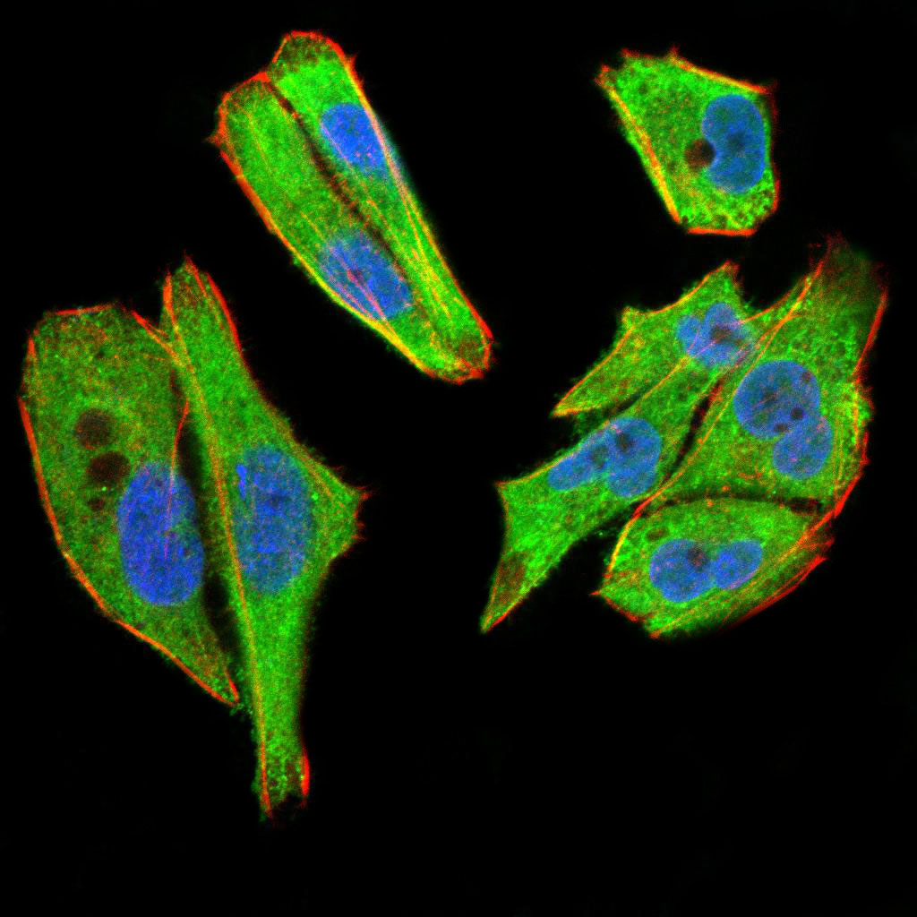
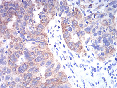
TBCC Antibody
Purified Mouse Monoclonal Antibody

- 产品详情
- 实验流程
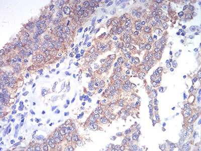
Application
| WB, IHC, ICC, E |
|---|---|
| Primary Accession | Q15814 |
| Reactivity | Human |
| Host | Mouse |
| Clonality | Monoclonal |
| Clone Names | 7G6H1 |
| Isotype | IgG1 |
| Calculated MW | 39248 Da |
| Description | Cofactor C is one of four proteins (cofactors A, D, E, and C) involved in the pathway leading to correctly folded beta-tubulin from folding intermediates. Cofactors A and D are believed to play a role in capturing and stabilizing beta-tubulin intermediates in a quasi-native confirmation. Cofactor E binds to the cofactor D/beta-tubulin complex; interaction with cofactor C then causes the release of beta-tubulin polypeptides that are committed to the native state. |
| Immunogen | Purified recombinant fragment of human *** (AA: 1-196) expressed in E. Coli. |
| Formulation | Purified antibody in PBS with 0.05% sodium azide |
| Gene ID | 6903 |
|---|---|
| Other Names | Tubulin-specific chaperone C, Tubulin-folding cofactor C, CFC, TBCC |
| Dilution | WB~~1/500 - 1/2000 IHC~~1/200 - 1/1000 ICC~~N/A E~~1/10000 |
| Storage | Maintain refrigerated at 2-8°C for up to 6 months. For long term storage store at -20°C in small aliquots to prevent freeze-thaw cycles. |
| Precautions | TBCC Antibody is for research use only and not for use in diagnostic or therapeutic procedures. |
| Name | TBCC |
|---|---|
| Function | Tubulin-folding protein; involved in the final step of the tubulin folding pathway. |
| Cellular Location | Cytoplasm. Note=Detected predominantly in the photoreceptor connecting cilium |
| Tissue Location | Expressed in the retina. Expressed in the rod and cone photoreceptors, extending from the inner segments (IS), through the outer nuclear layer (ONL) and into the synapses in the outer plexiform layer (OPL). Strongly expressed to the photoreceptor connecting cilium at the tips of the IS (at protein level) |
Research Areas
For Research Use Only. Not For Use In Diagnostic Procedures.
Application Protocols
Provided below are standard protocols that you may find useful for product applications.
REFERENCES
1.PLoS One. 2011;6(10):e25912.2.BMC Cancer. 2010 Apr 12;10:135.
终于等到您。ABCEPTA(百远生物)抗体产品。
点击下方“我要评价 ”按钮提交您的反馈信息,您的反馈和评价是我们最宝贵的财富之一,
我们将在1-3个工作日内处理您的反馈信息。
如有疑问,联系:0512-88856768 tech-china@abcepta.com.


