


 癌症的基本特征包括细胞增殖、血管生成、迁移、凋亡逃避机制和细胞永生等。找到癌症发生过程中这些通路的关键标记物和对应的抗体用于检测至关重要。
 为您推荐一个泛素化位点预测神器——泛素化分析工具,可以为您的蛋白的泛素化位点作出预测和评分。
 细胞自噬受体图形绘图工具为你的蛋白的细胞受体结合位点作出预测和评分,识别结合到自噬通路中的蛋白是非常重要的,便于让我们理解自噬在正常生理、病理过程中的作用,如发育、细胞分化、神经退化性疾病、压力条件下、感染和癌症。
SDC1 Antibody
Purified Mouse Monoclonal Antibody
- 产品详情
- 实验流程
- 背景知识
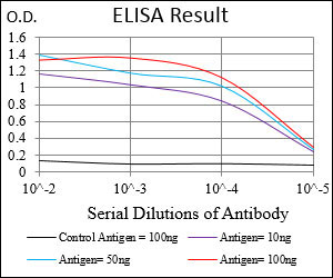
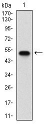
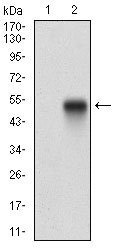
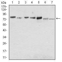
Application
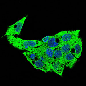
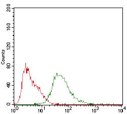
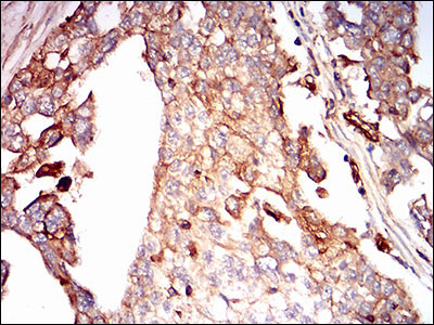
| WB, IHC, FC, ICC, E |
|---|---|
| Primary Accession | P18827 |
| Reactivity | Human, Mouse |
| Host | Mouse |
| Clonality | Monoclonal |
| Clone Names | 1A3H4 |
| Isotype | IgG1 |
| Calculated MW | 32462 Da |
| Description | The protein encoded by this gene is a transmembrane (type I) heparan sulfate proteoglycan and is a member of the syndecan proteoglycan family. The syndecans mediate cell binding, cell signaling, and cytoskeletal organization and syndecan receptors are required for internalization of the HIV-1 tat protein. The syndecan-1 protein functions as an integral membrane protein and participates in cell proliferation, cell migration and cell-matrix interactions via its receptor for extracellular matrix proteins. Altered syndecan-1 expression has been detected in several different tumor types. While several transcript variants may exist for this gene, the full-length natures of only two have been described to date. These two represent the major variants of this gene and encode the same protein. |
| Immunogen | Purified recombinant fragment of human SDC1 (AA: 28-171) expressed in E. Coli. |
| Formulation | Purified antibody in PBS with 0.05% sodium azide. |
| Other Names | Syndecan-1, SYND1, CD138, SDC1, SDC |
|---|---|
| Dilution | WB~~1/500 - 1/2000 IHC~~1/200 - 1/1000 FC~~1/200 - 1/400 ICC~~N/A E~~1/10000 |
| Storage | Maintain refrigerated at 2-8°C for up to 6 months. For long term storage store at -20°C in small aliquots to prevent freeze-thaw cycles. |
| Precautions | SDC1 Antibody is for research use only and not for use in diagnostic or therapeutic procedures. |
For Research Use Only. Not For Use In Diagnostic Procedures.
Provided below are standard protocols that you may find useful for product applications.
BACKGROUND
The multi-pass membrane protein encoded by this gene belongs to the G-protein coupled receptor 3 family and GABA-B receptor subfamily. The GABA-B receptors inhibit neuronal activity through G protein-coupled second-messenger systems, which regulate the release of neurotransmitters, and the activity of ion channels and adenylyl cyclase. This receptor subunit forms an active heterodimeric complex with GABA-B receptor subunit 1, neither of which is effective on its own. Allelic variants of this gene have been associated with nicotine dependence. ; ;
REFERENCES
1. Am J Clin Pathol. 2012 Mar;137(3):423-8. 2. PLoS One. 2011;6(9):e25252.
终于等到您。ABCEPTA(百远生物)抗体产品。
点击下方“我要评价 ”按钮提交您的反馈信息,您的反馈和评价是我们最宝贵的财富之一,
我们将在1-3个工作日内处理您的反馈信息。
如有疑问,联系:0512-88856768 tech-china@abcepta.com.


