


 癌症的基本特征包括细胞增殖、血管生成、迁移、凋亡逃避机制和细胞永生等。找到癌症发生过程中这些通路的关键标记物和对应的抗体用于检测至关重要。
 为您推荐一个泛素化位点预测神器——泛素化分析工具,可以为您的蛋白的泛素化位点作出预测和评分。
 细胞自噬受体图形绘图工具为你的蛋白的细胞受体结合位点作出预测和评分,识别结合到自噬通路中的蛋白是非常重要的,便于让我们理解自噬在正常生理、病理过程中的作用,如发育、细胞分化、神经退化性疾病、压力条件下、感染和癌症。
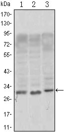
CD69 Antibody
Purified Mouse Monoclonal Antibody

- 产品详情
- 实验流程
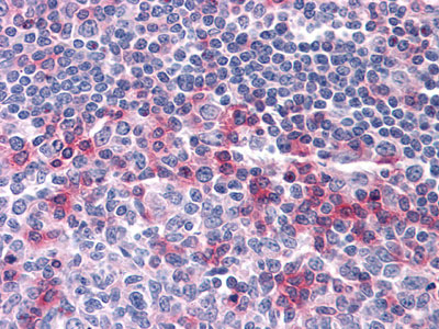
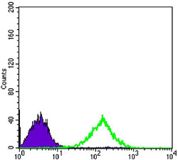
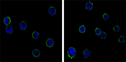
Application
| WB, IHC, FC, E |
|---|---|
| Primary Accession | Q07108 |
| Reactivity | Human |
| Host | Mouse |
| Clonality | Monoclonal |
| Clone Names | 8B6 |
| Isotype | IgG1 |
| Calculated MW | 22559 Da |
| Description | Involved in lymphocyte proliferation and functions as a signal transmitting receptor in lymphocytes, natural killer (NK) cells, andplatelets Subcellular location: Membrane, Single-pass type II membrane protein Tissue specificity: Expressed on the surface of activated T-cells, B-cells, natural killer cells, neutrophils, eosinophils, epidermal Langerhanscells and platelets Sequence similarities: Contains 1 C-type lectin domain. |
| Immunogen | Purified recombinant fragment of human CD69 expressed in E. Coli. |
| Formulation | Ascitic fluid containing 0.03% sodium azide. |
| Gene ID | 969 |
|---|---|
| Other Names | Early activation antigen CD69, Activation inducer molecule, AIM, BL-AC/P26, C-type lectin domain family 2 member C, EA1, Early T-cell activation antigen p60, GP32/28, Leukocyte surface antigen Leu-23, MLR-3, CD69, CD69, CLEC2C |
| Dilution | WB~~1/500 - 1/2000 IHC~~1/200 - 1/1000 FC~~1/200 - 1/400 E~~N/A |
| Storage | Maintain refrigerated at 2-8°C for up to 6 months. For long term storage store at -20°C in small aliquots to prevent freeze-thaw cycles. |
| Precautions | CD69 Antibody is for research use only and not for use in diagnostic or therapeutic procedures. |
| Name | CD69 |
|---|---|
| Synonyms | CLEC2C |
| Function | Transmembrane protein expressed mainly on T-cells resident in mucosa that plays an essential role in immune cell homeostasis. Rapidly expressed on the surface of platelets, T-lymphocytes and NK cells upon activation by various stimuli, such as antigen recognition or cytokine signaling, stimulates different signaling pathways in different cell types (PubMed:24752896, PubMed:26296369, PubMed:35930205). Negatively regulates Th17 cell differentiation through its carbohydrate dependent interaction with galectin-1/LGALS1 present on immature dendritic cells (PubMed:24752896). Association of CD69 cytoplasmic tail with the JAK3/STAT5 signaling pathway regulates the transcription of RORgamma/RORC and, consequently, differentiation toward the Th17 lineage (By similarity). Also acts via the S100A8/S100A9 complex present on peripheral blood mononuclear cells to promote the conversion of naive CD4 T-cells into regulatory T-cells (PubMed:26296369). Acts as an oxidized low-density lipoprotein (oxLDL) receptor in CD4 T- lymphocytes and negatively regulates the inflammatory response by inducing the expression of PDCD1 through the activation of NFAT (PubMed:35930205). Participates in adipose tissue-derived mesenchymal stem cells (ASCs)-mediated protection against P.aeruginosa infection. Mechanistically, specifically recognizes P.aeruginosa to promote ERK1 activation, followed by granulocyte-macrophage colony-stimulating factor (GM-CSF) and other inflammatory cytokines secretion (PubMed:34841721). In eosinophils, induces IL-10 production through the ERK1/2 pathway (By similarity). Negatively regulates the chemotactic responses of effector lymphocytes and dendritic cells (DCs) to sphingosine 1 phosphate/S1P by acting as a S1PR1 receptor agonist and facilitating the internalization and degradation of the receptor (PubMed:37039481). |
| Cellular Location | Cell membrane; Single-pass type II membrane protein |
| Tissue Location | Expressed on the surface of activated T-cells, B- cells, natural killer cells, neutrophils, eosinophils, epidermal Langerhans cells and platelets |
Research Areas
For Research Use Only. Not For Use In Diagnostic Procedures.
Application Protocols
Provided below are standard protocols that you may find useful for product applications.
REFERENCES
1. EMBO J. 1997 Feb 17;16(4):673-84. 2. Cell Immunol. 2002 Nov;220(1):20-9. 3. Arch Biochem Biophys. 2005 Jun 1;438(1):11-20.
终于等到您。ABCEPTA(百远生物)抗体产品。
点击下方“我要评价 ”按钮提交您的反馈信息,您的反馈和评价是我们最宝贵的财富之一,
我们将在1-3个工作日内处理您的反馈信息。
如有疑问,联系:0512-88856768 tech-china@abcepta.com.
¥ 1,500.00
Cat# AO1374a


