


 癌症的基本特征包括细胞增殖、血管生成、迁移、凋亡逃避机制和细胞永生等。找到癌症发生过程中这些通路的关键标记物和对应的抗体用于检测至关重要。
 为您推荐一个泛素化位点预测神器——泛素化分析工具,可以为您的蛋白的泛素化位点作出预测和评分。
 细胞自噬受体图形绘图工具为你的蛋白的细胞受体结合位点作出预测和评分,识别结合到自噬通路中的蛋白是非常重要的,便于让我们理解自噬在正常生理、病理过程中的作用,如发育、细胞分化、神经退化性疾病、压力条件下、感染和癌症。
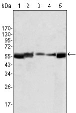
LYN Antibody
Purified Mouse Monoclonal Antibody
- 产品详情
- 实验流程
Application
| WB, E |
|---|---|
| Primary Accession | P07948 |
| Reactivity | Human |
| Host | Mouse |
| Clonality | Monoclonal |
| Clone Names | 2H8D7 |
| Isotype | IgG2b |
| Calculated MW | 58574 Da |
| Description | Lyn (also known as p53/56 Lyn) is a membrane-associated protein tyrosine kinase (PTK) mostly expressed in hemopoietic cells which is important in cellular signaling. It contains an SH2 and SH3 domain and has been found to be cleaved after activation of caspases in apoptosis. A member of the Src family of PTKs, there are two known isoforms for Lyn which plays an indispensable role in the Fc epsilon RI (Fcer1) and the B-cell IgM receptor signaling pathway and is essential for Syk activation and Lat phosphorylation after Fcer1 aggregation and can also phosphor-ylate Tec on multiple residues. Lyn can also be regulated by IL-2 and IL-3.Lyn is a member of the src family of non-receptor protein tyrosine kinases that is predominantly expressed in haematopoietic tissues. Like all members of the src family, lyn is thought to participate in signal transduction from cell surface receptors that lack intrinsic tyrosine kinase activity. It is associated with a number of cell surface receptors including the B cell antigen receptor and immunoglobulin E receptor (FceRI). |
| Immunogen | Purified recombinant fragment of LYN expressed in E. Coli. |
| Formulation | Ascitic fluid containing 0.03% sodium azide. |
| Gene ID | 4067 |
|---|---|
| Other Names | Tyrosine-protein kinase Lyn, 2.7.10.2, Lck/Yes-related novel protein tyrosine kinase, V-yes-1 Yamaguchi sarcoma viral related oncogene homolog, p53Lyn, p56Lyn, LYN, JTK8 |
| Dilution | WB~~1/500 - 1/2000 E~~N/A |
| Storage | Maintain refrigerated at 2-8°C for up to 6 months. For long term storage store at -20°C in small aliquots to prevent freeze-thaw cycles. |
| Precautions | LYN Antibody is for research use only and not for use in diagnostic or therapeutic procedures. |
| Name | LYN |
|---|---|
| Synonyms | JTK8 |
| Function | Non-receptor tyrosine-protein kinase that transmits signals from cell surface receptors and plays an important role in the regulation of innate and adaptive immune responses, hematopoiesis, responses to growth factors and cytokines, integrin signaling, but also responses to DNA damage and genotoxic agents. Functions primarily as negative regulator, but can also function as activator, depending on the context. Required for the initiation of the B-cell response, but also for its down-regulation and termination. Plays an important role in the regulation of B-cell differentiation, proliferation, survival and apoptosis, and is important for immune self-tolerance. Acts downstream of several immune receptors, including the B-cell receptor, CD79A, CD79B, CD5, CD19, CD22, FCER1, FCGR2, FCGR1A, TLR2 and TLR4. Plays a role in the inflammatory response to bacterial lipopolysaccharide. Mediates the responses to cytokines and growth factors in hematopoietic progenitors, platelets, erythrocytes, and in mature myeloid cells, such as dendritic cells, neutrophils and eosinophils. Acts downstream of EPOR, KIT, MPL, the chemokine receptor CXCR4, as well as the receptors for IL3, IL5 and CSF2. Plays an important role in integrin signaling. Regulates cell proliferation, survival, differentiation, migration, adhesion, degranulation, and cytokine release. Involved in the regulation of endothelial activation, neutrophil adhesion and transendothelial migration (PubMed:36932076). Down-regulates signaling pathways by phosphorylation of immunoreceptor tyrosine-based inhibitory motifs (ITIM), that then serve as binding sites for phosphatases, such as PTPN6/SHP-1, PTPN11/SHP-2 and INPP5D/SHIP-1, that modulate signaling by dephosphorylation of kinases and their substrates. Phosphorylates LIME1 in response to CD22 activation. Phosphorylates BTK, CBL, CD5, CD19, CD72, CD79A, CD79B, CSF2RB, DOK1, HCLS1, LILRB3/PIR-B, MS4A2/FCER1B, SYK and TEC. Promotes phosphorylation of SIRPA, PTPN6/SHP-1, PTPN11/SHP-2 and INPP5D/SHIP-1. Mediates phosphorylation of the BCR-ABL fusion protein. Required for rapid phosphorylation of FER in response to FCER1 activation. Mediates KIT phosphorylation. Acts as an effector of EPOR (erythropoietin receptor) in controlling KIT expression and may play a role in erythroid differentiation during the switch between proliferation and maturation. Depending on the context, activates or inhibits several signaling cascades. Regulates phosphatidylinositol 3-kinase activity and AKT1 activation. Regulates activation of the MAP kinase signaling cascade, including activation of MAP2K1/MEK1, MAPK1/ERK2, MAPK3/ERK1, MAPK8/JNK1 and MAPK9/JNK2. Mediates activation of STAT5A and/or STAT5B. Phosphorylates LPXN on 'Tyr-72'. Kinase activity facilitates TLR4-TLR6 heterodimerization and signal initiation. Phosphorylates SCIMP on 'Tyr- 107'; this enhances binding of SCIMP to TLR4, promoting the phosphorylation of TLR4, and a selective cytokine response to lipopolysaccharide in macrophages (By similarity). Phosphorylates CLNK (By similarity). Phosphorylates BCAR1/CAS and NEDD9/HEF1 (PubMed:9020138). |
| Cellular Location | Cell membrane. Nucleus. Cytoplasm. Cytoplasm, perinuclear region. Golgi apparatus. Membrane; Lipid- anchor. Note=Accumulates in the nucleus by inhibition of CRM1-mediated nuclear export. Nuclear accumulation is increased by inhibition of its kinase activity. The trafficking from the Golgi apparatus to the plasma membrane occurs in a kinase domain-dependent but kinase activity independent manner and is mediated by exocytic vesicular transport. Detected on plasma membrane lipid rafts |
| Tissue Location | Detected in monocytes (at protein level). Detected in placenta, and in fetal brain, lung, liver and kidney. Widely expressed in a variety of organs, tissues, and cell types such as epidermoid, hematopoietic, and neuronal cells. Expressed in primary neuroblastoma tumors. |
Research Areas
For Research Use Only. Not For Use In Diagnostic Procedures.
Application Protocols
Provided below are standard protocols that you may find useful for product applications.
REFERENCES
1. Sakaguchi, A.Y.,et al.Genet. 34: 175. 2. Hibbs, M.L.,et al.Biol. 29: 397-400. 3. Williams, J.C.,et al.Trends Biochem. Sci. 23: 179-184.
终于等到您。ABCEPTA(百远生物)抗体产品。
点击下方“我要评价 ”按钮提交您的反馈信息,您的反馈和评价是我们最宝贵的财富之一,
我们将在1-3个工作日内处理您的反馈信息。
如有疑问,联系:0512-88856768 tech-china@abcepta.com.
¥ 1,500.00
Cat# AO1097a


