


 癌症的基本特征包括细胞增殖、血管生成、迁移、凋亡逃避机制和细胞永生等。找到癌症发生过程中这些通路的关键标记物和对应的抗体用于检测至关重要。
 为您推荐一个泛素化位点预测神器——泛素化分析工具,可以为您的蛋白的泛素化位点作出预测和评分。
 细胞自噬受体图形绘图工具为你的蛋白的细胞受体结合位点作出预测和评分,识别结合到自噬通路中的蛋白是非常重要的,便于让我们理解自噬在正常生理、病理过程中的作用,如发育、细胞分化、神经退化性疾病、压力条件下、感染和癌症。
C-Kit Antibody
Purified Mouse Monoclonal Antibody
- 产品详情
- 实验流程
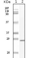
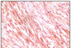
Application
| WB, IHC, E |
|---|---|
| Primary Accession | P10721 |
| Reactivity | Human |
| Host | Mouse |
| Clonality | Monoclonal |
| Clone Names | 8D7 |
| Isotype | IgG1 |
| Calculated MW | 109865 Da |
| Description | C-kit (CD117, 145kDa) functions as a tyrosine kinase receptor which becomes activated upon binding of its ligand SCF (stem-cell factor), the C-kit gene encodes the human homolog of the proto-oncogene c-kit. which was first identified as the cellular homolog of the feline sarcoma viral oncogene v-kit. KIT is a type 3 transmembrane receptor for MGF (mast cell growth factor). Mutations in KIT are associated with gastrointestinal stromal tumors, mast cell disease, acute myelogenous lukemia, and piebaldism. |
| Immunogen | Purified recombinant fragment of C-kit expressed in E. Coli. |
| Formulation | Ascitic fluid containing 0.03% sodium azide. |
| Gene ID | 3815 |
|---|---|
| Other Names | Mast/stem cell growth factor receptor Kit, SCFR, 2.7.10.1, Piebald trait protein, PBT, Proto-oncogene c-Kit, Tyrosine-protein kinase Kit, p145 c-kit, v-kit Hardy-Zuckerman 4 feline sarcoma viral oncogene homolog, CD117, KIT, SCFR |
| Dilution | WB~~1/500 - 1/2000 IHC~~1/200 - 1/1000 E~~N/A |
| Storage | Maintain refrigerated at 2-8°C for up to 6 months. For long term storage store at -20°C in small aliquots to prevent freeze-thaw cycles. |
| Precautions | C-Kit Antibody is for research use only and not for use in diagnostic or therapeutic procedures. |
| Name | KIT |
|---|---|
| Synonyms | SCFR |
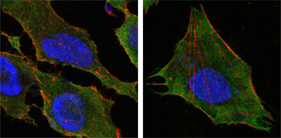
| Function | Tyrosine-protein kinase that acts as a cell-surface receptor for the cytokine KITLG/SCF and plays an essential role in the regulation of cell survival and proliferation, hematopoiesis, stem cell maintenance, gametogenesis, mast cell development, migration and function, and in melanogenesis. In response to KITLG/SCF binding, KIT can activate several signaling pathways. Phosphorylates PIK3R1, PLCG1, SH2B2/APS and CBL. Activates the AKT1 signaling pathway by phosphorylation of PIK3R1, the regulatory subunit of phosphatidylinositol 3-kinase. Activated KIT also transmits signals via GRB2 and activation of RAS, RAF1 and the MAP kinases MAPK1/ERK2 and/or MAPK3/ERK1. Promotes activation of STAT family members STAT1, STAT3, STAT5A and STAT5B. Activation of PLCG1 leads to the production of the cellular signaling molecules diacylglycerol and inositol 1,4,5- trisphosphate. KIT signaling is modulated by protein phosphatases, and by rapid internalization and degradation of the receptor. Activated KIT promotes phosphorylation of the protein phosphatases PTPN6/SHP-1 and PTPRU, and of the transcription factors STAT1, STAT3, STAT5A and STAT5B. Promotes phosphorylation of PIK3R1, CBL, CRK (isoform Crk-II), LYN, MAPK1/ERK2 and/or MAPK3/ERK1, PLCG1, SRC and SHC1. |
| Cellular Location | [Isoform 1]: Cell membrane; Single-pass type I membrane protein [Isoform 3]: Cytoplasm. Note=Detected in the cytoplasm of spermatozoa, especially in the equatorial and subacrosomal region of the sperm head. |
| Tissue Location | [Isoform 3]: In testis, detected in spermatogonia in the basal layer and in interstitial Leydig cells but not in Sertoli cells or spermatocytes inside the seminiferous tubules (at protein level) (PubMed:20601678). Expression is maintained in ejaculated spermatozoa (at protein level) (PubMed:20601678) |
Research Areas
For Research Use Only. Not For Use In Diagnostic Procedures.
Application Protocols
Provided below are standard protocols that you may find useful for product applications.
REFERENCES
1. Mojica WD et.alHistopathology. 2005 Nov;47(5):517-22. 2. Tong WD et.alInt J Colorectal Dis. 2005 Jul;20(4):363-7. Epub 2005 Feb 2. 3. Nakai Y et.alBiochem Biophys Res Commun. 2005 Nov 11;337(1):289-96.
终于等到您。ABCEPTA(百远生物)抗体产品。
点击下方“我要评价 ”按钮提交您的反馈信息,您的反馈和评价是我们最宝贵的财富之一,
我们将在1-3个工作日内处理您的反馈信息。
如有疑问,联系:0512-88856768 tech-china@abcepta.com.
¥ 1,500.00
Cat# AO1095a


