


 癌症的基本特征包括细胞增殖、血管生成、迁移、凋亡逃避机制和细胞永生等。找到癌症发生过程中这些通路的关键标记物和对应的抗体用于检测至关重要。
 为您推荐一个泛素化位点预测神器——泛素化分析工具,可以为您的蛋白的泛素化位点作出预测和评分。
 细胞自噬受体图形绘图工具为你的蛋白的细胞受体结合位点作出预测和评分,识别结合到自噬通路中的蛋白是非常重要的,便于让我们理解自噬在正常生理、病理过程中的作用,如发育、细胞分化、神经退化性疾病、压力条件下、感染和癌症。
HINT1 Antibody
Purified Mouse Monoclonal Antibody (Mab)

- 产品详情
- 实验流程
- 背景知识
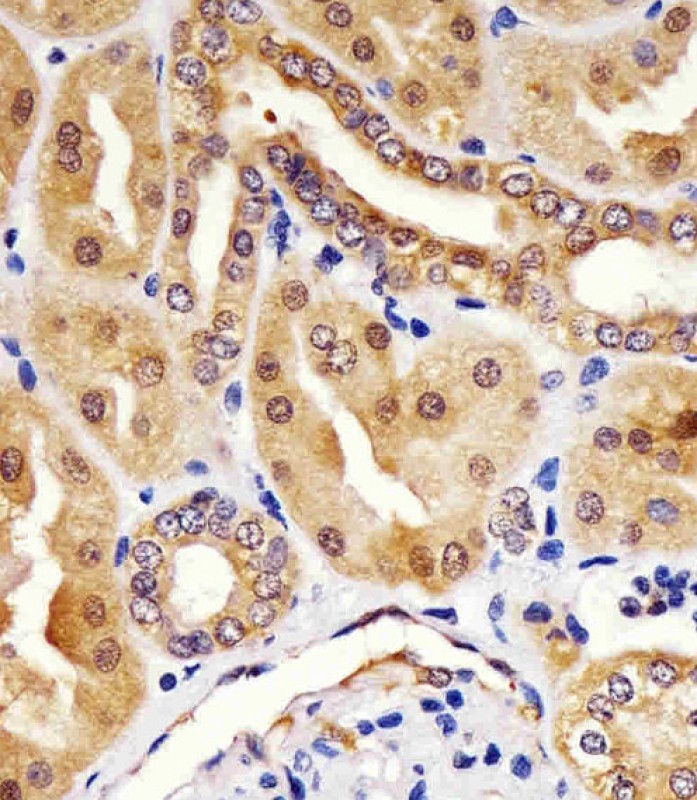
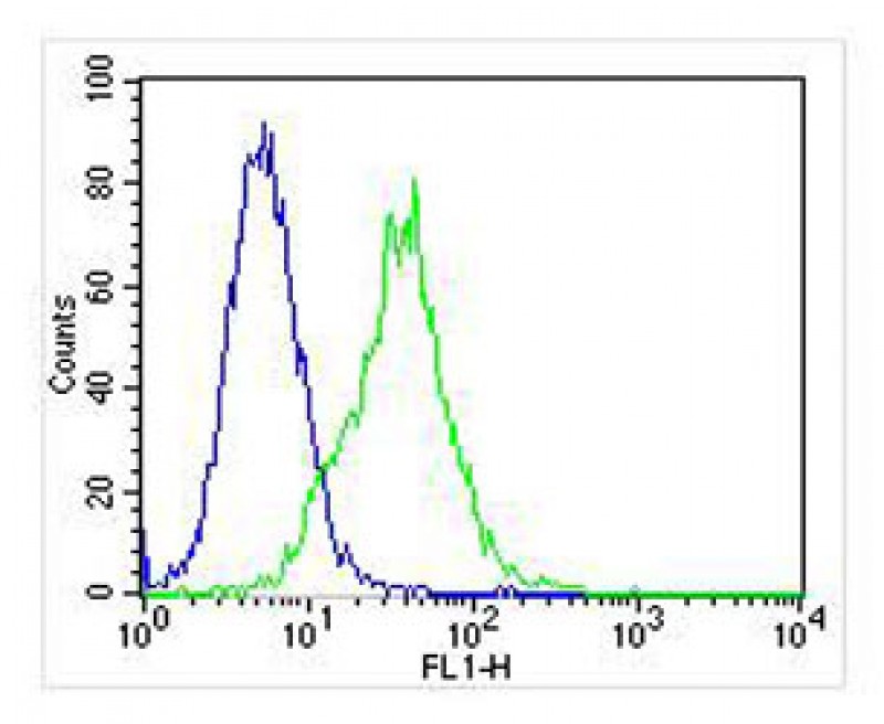
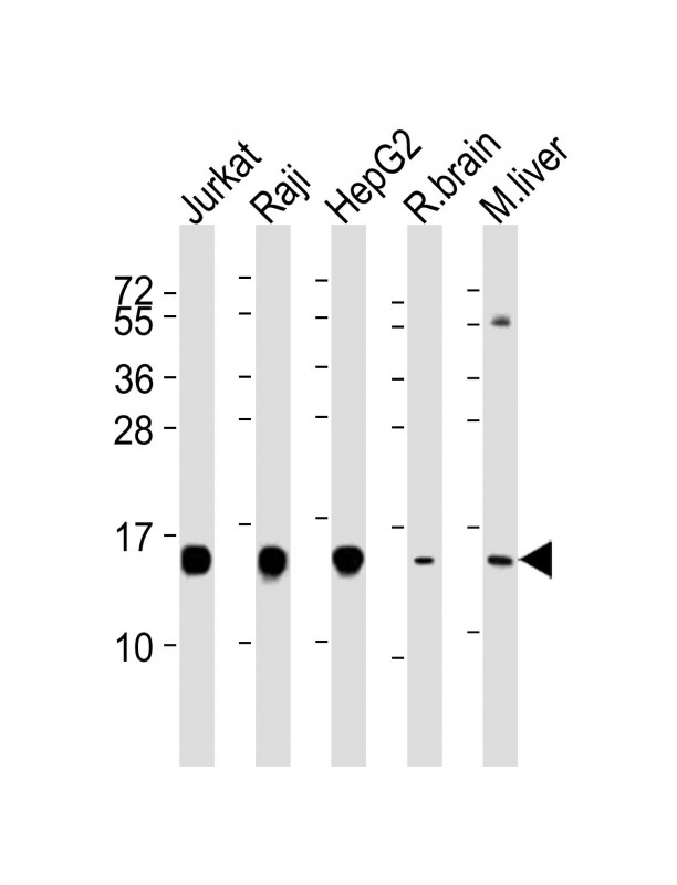
Application
| WB, FC, IHC-P, IF, E |
|---|---|
| Primary Accession | P49773 |
| Reactivity | Human, Mouse, Rat |
| Host | Mouse |
| Clonality | monoclonal |
| Isotype | IgG1,k |
| Clone Names | 1500CT836.13.93 |
| Calculated MW | 13802 Da |
| Gene ID | 3094 |
|---|---|
| Other Names | Histidine triad nucleotide-binding protein 1, 3---, Adenosine 5'-monophosphoramidase, Protein kinase C inhibitor 1, Protein kinase C-interacting protein 1, PKCI-1, HINT1, HINT, PKCI1, PRKCNH1 |
| Target/Specificity | This HINT1 antibody is generated from a mouse immunized with a recombinant protein of human HINT1. |
| Dilution | WB~~1:4000 FC~~1:25 IHC-P~~1:100~500 IF~~1:25 E~~Use at an assay dependent concentration. |
| Format | Purified monoclonal antibody supplied in PBS with 0.09% (W/V) sodium azide. This antibody is purified through a protein G column, followed by dialysis against PBS. |
| Storage | Maintain refrigerated at 2-8°C for up to 2 weeks. For long term storage store at -20°C in small aliquots to prevent freeze-thaw cycles. |
| Precautions | HINT1 Antibody is for research use only and not for use in diagnostic or therapeutic procedures. |
| Name | HINT1 |
|---|---|
| Synonyms | HINT, PKCI1, PRKCNH1 |
| Function | Exhibits adenosine 5'-monophosphoramidase activity, hydrolyzing purine nucleotide phosphoramidates with a single phosphate group such as adenosine 5'monophosphoramidate (AMP-NH2) to yield AMP and NH2 (PubMed:15703176, PubMed:16835243, PubMed:17217311, PubMed:17337452, PubMed:22329685, PubMed:23614568, PubMed:28691797, PubMed:29787766, PubMed:31990367). Hydrolyzes adenosine 5'monophosphomorpholidate (AMP-morpholidate) and guanosine 5'monophosphomorpholidate (GMP-morpholidate) (PubMed:15703176, PubMed:16835243). Hydrolyzes lysyl-AMP (AMP-N-epsilon-(N-alpha-acetyl lysine methyl ester)) generated by lysine tRNA ligase, as well as Met- AMP, His-AMP and Asp-AMP, lysyl-GMP (GMP-N-epsilon-(N-alpha-acetyl lysine methyl ester)) and AMP-N-alanine methyl ester (PubMed:15703176, PubMed:17337452, PubMed:22329685). Hydrolyzes 3-indolepropionic acyl- adenylate, tryptamine adenosine phosphoramidate monoester and other fluorogenic purine nucleoside tryptamine phosphoramidates in vitro (PubMed:17217311, PubMed:17337452, PubMed:23614568, PubMed:28691797, PubMed:29787766, PubMed:31990367). Can also convert adenosine 5'-O- phosphorothioate and guanosine 5'-O-phosphorothioate to the corresponding nucleoside 5'-O-phosphates with concomitant release of hydrogen sulfide (PubMed:30772266). In addition, functions as scaffolding protein that modulates transcriptional activation by the LEF1/TCF1-CTNNB1 complex and by the complex formed with MITF and CTNNB1 (PubMed:16014379, PubMed:22647378). Modulates p53/TP53 levels and p53/TP53-mediated apoptosis (PubMed:16835243). Modulates proteasomal degradation of target proteins by the SCF (SKP2-CUL1-F-box protein) E3 ubiquitin-protein ligase complex (PubMed:19112177). Also exhibits SUMO- specific isopeptidase activity, deconjugating SUMO1 from RGS17 (PubMed:31088288). Deconjugates SUMO1 from RANGAP1 (By similarity). |
| Cellular Location | Cytoplasm. Nucleus. Note=Interaction with CDK7 leads to a more nuclear localization. |
| Tissue Location | Widely expressed. |
For Research Use Only. Not For Use In Diagnostic Procedures.
Provided below are standard protocols that you may find useful for product applications.
BACKGROUND
Hydrolyzes purine nucleotide phosphoramidates with a single phosphate group, including adenosine 5'monophosphoramidate (AMP-NH2), adenosine 5'monophosphomorpholidate (AMP-morpholidate) and guanosine 5'monophosphomorpholidate (GMP-morpholidate). Hydrolyzes lysyl-AMP (AMP-N-epsilon-(N-alpha-acetyl lysine methyl ester)) generated by lysine tRNA ligase, as well as Met-AMP, His- AMP and Asp-AMP, lysyl-GMP (GMP-N-epsilon-(N-alpha-acetyl lysine methyl ester)) and AMP-N-alanine methyl ester. Can also convert adenosine 5'-O-phosphorothioate and guanosine 5'-O- phosphorothioate to the corresponding nucleoside 5'-O-phosphates with concomitant release of hydrogen sulfide. In addition, functions as scaffolding protein that modulates transcriptional activation by the LEF1/TCF1-CTNNB1 complex and by the complex formed with MITF and CTNNB1. Modulates p53/TP53 levels and p53/TP53-mediated apoptosis. Modulates proteasomal degradation of target proteins by the SCF (SKP2-CUL1-F-box protein) E3 ubiquitin- protein ligase complex.
REFERENCES
Brzoska P.M.,et al.Genomics 36:151-156(1996).
Brzoska P.M.,et al.Proc. Natl. Acad. Sci. U.S.A. 92:7824-7828(1995).
Ota T.,et al.Nat. Genet. 36:40-45(2004).
Ebert L.,et al.Submitted (JUN-2004) to the EMBL/GenBank/DDBJ databases.
Lima C.D.,et al.Proc. Natl. Acad. Sci. U.S.A. 93:5357-5362(1996).
终于等到您。ABCEPTA(百远生物)抗体产品。
点击下方“我要评价 ”按钮提交您的反馈信息,您的反馈和评价是我们最宝贵的财富之一,
我们将在1-3个工作日内处理您的反馈信息。
如有疑问,联系:0512-88856768 tech-china@abcepta.com.


