


 癌症的基本特征包括细胞增殖、血管生成、迁移、凋亡逃避机制和细胞永生等。找到癌症发生过程中这些通路的关键标记物和对应的抗体用于检测至关重要。
 为您推荐一个泛素化位点预测神器——泛素化分析工具,可以为您的蛋白的泛素化位点作出预测和评分。
 细胞自噬受体图形绘图工具为你的蛋白的细胞受体结合位点作出预测和评分,识别结合到自噬通路中的蛋白是非常重要的,便于让我们理解自噬在正常生理、病理过程中的作用,如发育、细胞分化、神经退化性疾病、压力条件下、感染和癌症。
VIME Antibody
Mouse Monoclonal Antibody (Mab)
- 产品详情
- 实验流程
- 背景知识
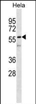
Application
| WB, E |
|---|---|
| Primary Accession | P08670 |
| Other Accession | NP_003371.2 |
| Reactivity | Human |
| Host | Mouse |
| Clonality | Monoclonal |
| Isotype | IgM,k |
| Clone Names | 280CT3.4.6 |
| Calculated MW | 53652 Da |
| Gene ID | 7431 |
|---|---|
| Other Names | Vimentin, VIM |
| Target/Specificity | This VIME monoclonal antibody is generated from mouse immunized with VIME recombinant protein. |
| Dilution | WB~~1:500~1000 E~~Use at an assay dependent concentration. |
| Format | Purified monoclonal antibody supplied in PBS with 0.09% (W/V) sodium azide. This antibody is prepared by Euglobin precipitation followed by dialysis against PBS. |
| Storage | Maintain refrigerated at 2-8°C for up to 2 weeks. For long term storage store at -20°C in small aliquots to prevent freeze-thaw cycles. |
| Precautions | VIME Antibody is for research use only and not for use in diagnostic or therapeutic procedures. |
| Name | VIM (HGNC:12692) |
|---|---|
| Function | Vimentins are class-III intermediate filaments found in various non-epithelial cells, especially mesenchymal cells. Vimentin is attached to the nucleus, endoplasmic reticulum, and mitochondria, either laterally or terminally. Plays a role in cell directional movement, orientation, cell sheet organization and Golgi complex polarization at the cell migration front (By similarity). Protects SCRIB from proteasomal degradation and facilitates its localization to intermediate filaments in a cell contact-mediated manner (By similarity). May promote axon outgrowth and motor fiber repair via DSP- mediated recruitment to outgrowth tips (By similarity). |
| Cellular Location | Cytoplasm. Cytoplasm, cytoskeleton. Nucleus matrix {ECO:0000250|UniProtKB:P31000}. Cell membrane {ECO:0000250|UniProtKB:P20152}. Cell projection, axon {ECO:0000250|UniProtKB:P20152} |
| Tissue Location | Highly expressed in fibroblasts, some expression in T- and B-lymphocytes, and little or no expression in Burkitt's lymphoma cell lines. Expressed in many hormone-independent mammary carcinoma cell lines. |
For Research Use Only. Not For Use In Diagnostic Procedures.
Provided below are standard protocols that you may find useful for product applications.
BACKGROUND
This gene encodes a member of the intermediate filament family. Intermediate filamentents, along with microtubules and actin microfilaments, make up the cytoskeleton. The protein encoded by this gene is responsible for maintaining cell shape, integrity of the cytoplasm, and stabilizing cytoskeletal interactions. It is also involved in the immune response, and controls the transport of low-density lipoprotein (LDL)-derived cholesterol from a lysosome to the site of esterification. It functions as an organizer of a number of critical proteins involved in attachment, migration, and cell signaling. Mutations in this gene causes a dominant, pulverulent cataract.
REFERENCES
Kers, J., et al. Transplantation 90(5):502-509(2010)
Pinheiro, A.P., et al. Am. J. Med. Genet. B Neuropsychiatr. Genet. 153B (5), 1070-1080 (2010) :
Korita, P.V., et al. Anticancer Res. 30(6):2279-2285(2010)
Martins-de-Souza, D., et al. J Psychiatr Res (2010) In press :
Li, M., et al. J. Exp. Clin. Cancer Res. 29, 109 (2010) :
终于等到您。ABCEPTA(百远生物)抗体产品。
点击下方“我要评价 ”按钮提交您的反馈信息,您的反馈和评价是我们最宝贵的财富之一,
我们将在1-3个工作日内处理您的反馈信息。
如有疑问,联系:0512-88856768 tech-china@abcepta.com.


