


 癌症的基本特征包括细胞增殖、血管生成、迁移、凋亡逃避机制和细胞永生等。找到癌症发生过程中这些通路的关键标记物和对应的抗体用于检测至关重要。
 为您推荐一个泛素化位点预测神器——泛素化分析工具,可以为您的蛋白的泛素化位点作出预测和评分。
 细胞自噬受体图形绘图工具为你的蛋白的细胞受体结合位点作出预测和评分,识别结合到自噬通路中的蛋白是非常重要的,便于让我们理解自噬在正常生理、病理过程中的作用,如发育、细胞分化、神经退化性疾病、压力条件下、感染和癌症。
BOK Antibody - N-terminal region
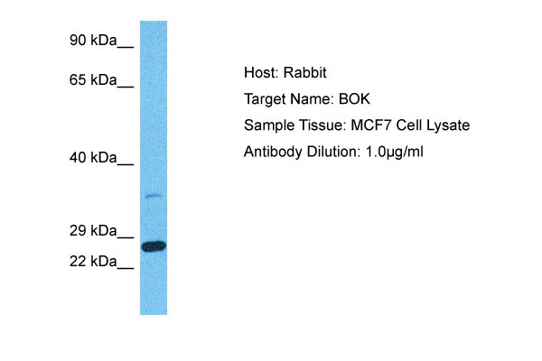
Rabbit Polyclonal Antibody
- 产品详情
- 实验流程
Application
| WB |
|---|---|
| Primary Accession | Q9UMX3 |
| Reactivity | Human |
| Predicted | Human, Mouse, Rat, Zebrafish, Bovine |
| Host | Rabbit |
| Clonality | Polyclonal |
| Calculated MW | 23280 Da |
| Gene ID | 666 |
|---|---|
| Alias Symbol | BOK, BCL2L9, |
| Other Names | Bcl-2-related ovarian killer protein, hBOK, Bcl-2-like protein 9, Bcl2-L-9, BOK, BCL2L9 |
| Format | Liquid. Purified antibody supplied in 1x PBS buffer with 0.09% (w/v) sodium azide and 2% sucrose. |
| Reconstitution & Storage | Add 50 &mu, l of distilled water. Final Anti-BOK antibody concentration is 1 mg/ml in PBS buffer with 2% sucrose. For longer periods of storage, store at -20°C. Avoid repeat freeze-thaw cycles. |
| Precautions | BOK Antibody - N-terminal region is for research use only and not for use in diagnostic or therapeutic procedures. |
| Name | BOK (HGNC:1087) |
|---|---|
| Synonyms | BCL2L9 |
| Function | [Isoform 1]: Apoptosis regulator that functions through different apoptotic signaling pathways (PubMed:15102863, PubMed:20673843, PubMed:27076518). Plays a roles as pro-apoptotic protein that positively regulates intrinsic apoptotic process in a BAX- and BAK1-dependent manner or in a BAX- and BAK1-independent manner (PubMed:15102863, PubMed:27076518). In response to endoplasmic reticulum stress promotes mitochondrial apoptosis through downstream BAX/BAK1 activation and positive regulation of PERK-mediated unfolded protein response (By similarity). Activates apoptosis independently of heterodimerization with survival-promoting BCL2 and BCL2L1 through induction of mitochondrial outer membrane permeabilization, in a BAX- and BAK1-independent manner, in response to inhibition of ERAD- proteasome degradation system, resulting in cytochrome c release (PubMed:27076518). In response to DNA damage, mediates intrinsic apoptotic process in a TP53-dependent manner (PubMed:15102863). Plays a role in granulosa cell apoptosis by CASP3 activation (PubMed:20673843). Plays a roles as anti-apoptotic protein during neuronal apoptotic process, by negatively regulating poly ADP-ribose polymerase-dependent cell death through regulation of neuronal calcium homeostasis and mitochondrial bioenergetics in response to NMDA excitation (By similarity). In addition to its role in apoptosis, may regulate trophoblast cell proliferation during the early stages of placental development, by acting on G1/S transition through regulation of CCNE1 expression (PubMed:19942931). May also play a role as an inducer of autophagy by disrupting interaction between MCL1 and BECN1 (PubMed:24113155). |
| Cellular Location | [Isoform 1]: Mitochondrion membrane {ECO:0000250|UniProtKB:O35425}; Single-pass membrane protein {ECO:0000250|UniProtKB:O35425}. Endoplasmic reticulum membrane; Single-pass membrane protein {ECO:0000250|UniProtKB:O35425}. Mitochondrion inner membrane. Cytoplasm. Nucleus. Mitochondrion. Endoplasmic reticulum. Mitochondrion outer membrane. Early endosome membrane {ECO:0000250|UniProtKB:O35425}. Recycling endosome membrane {ECO:0000250|UniProtKB:O35425}. Nucleus outer membrane {ECO:0000250|UniProtKB:O35425}. Golgi apparatus, cis-Golgi network membrane {ECO:0000250|UniProtKB:O35425}. Golgi apparatus, trans-Golgi network membrane {ECO:0000250|UniProtKB:O35425}. Membrane. Note=Nuclear and cytoplasmic compartments in the early stages of apoptosis and during apoptosis it associates with mitochondria (PubMed:19942931). In healthy cells, associates loosely with the membrane in a hit-and-run mode. The insertion and accumulation on membranes is enhanced through the activity of death signals, resulting in the integration of the membrane-bound protein into the membrane (PubMed:15868100). The transmembrane domain controls subcellular localization; constitutes a tail-anchor. Localizes in early and late endosome upon blocking of apoptosis. Must localize to the mitochondria to induce mitochondrial outer membrane permeabilization and apoptosis (By similarity) {ECO:0000250|UniProtKB:O35425, ECO:0000269|PubMed:15868100, ECO:0000269|PubMed:19942931} |
| Tissue Location | Expressed mainly in oocytes; weak expression in granulosa cells of the developing follicles. In adult human ovaries, expressed in granulosa cells at all follicular stages, but expression in primordial/primary follicles granulosa cell is stronger than in secondary and antral follicles. |
Research Areas
For Research Use Only. Not For Use In Diagnostic Procedures.
Application Protocols
Provided below are standard protocols that you may find useful for product applications.
REFERENCES
Zhang H.,et al.FEBS Lett. 480:311-313(2000).
Kalnine N.,et al.Submitted (MAY-2003) to the EMBL/GenBank/DDBJ databases.
Inohara N.,et al.J. Biol. Chem. 273:8705-8710(1998).
Mayya V.,et al.Sci. Signal. 2:RA46-RA46(2009).
Echeverry N.,et al.Cell Death Differ. 20:785-799(2013).
终于等到您。ABCEPTA(百远生物)抗体产品。
点击下方“我要评价 ”按钮提交您的反馈信息,您的反馈和评价是我们最宝贵的财富之一,
我们将在1-3个工作日内处理您的反馈信息。
如有疑问,联系:0512-88856768 tech-china@abcepta.com.


