


 癌症的基本特征包括细胞增殖、血管生成、迁移、凋亡逃避机制和细胞永生等。找到癌症发生过程中这些通路的关键标记物和对应的抗体用于检测至关重要。
 为您推荐一个泛素化位点预测神器——泛素化分析工具,可以为您的蛋白的泛素化位点作出预测和评分。
 细胞自噬受体图形绘图工具为你的蛋白的细胞受体结合位点作出预测和评分,识别结合到自噬通路中的蛋白是非常重要的,便于让我们理解自噬在正常生理、病理过程中的作用,如发育、细胞分化、神经退化性疾病、压力条件下、感染和癌症。
ADC antibody - middle region
Rabbit Polyclonal Antibody
- 产品详情
- 实验流程
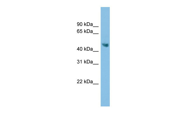
Application
| WB |
|---|---|
| Primary Accession | Q96A70 |
| Other Accession | NM_052998, NP_443724 |
| Reactivity | Human, Mouse, Rat, Rabbit, Pig, Dog, Guinea Pig, Horse, Bovine |
| Predicted | Human, Mouse, Rat, Rabbit, Pig, Dog, Horse, Bovine |
| Host | Rabbit |
| Clonality | Polyclonal |
| Calculated MW | 49980 Da |
| Gene ID | 113451 |
|---|---|
| Alias Symbol | AZI2, KIAA1945, ODC-p, ODC1L |
| Other Names | Antizyme inhibitor 2, AzI2, Arginine decarboxylase, ADC, ARGDC, Ornithine decarboxylase-like protein, ODC-like protein, ornithine decarboxylase paralog, ODC-p, AZIN2, ADC, KIAA1945, ODCP |
| Format | Liquid. Purified antibody supplied in 1x PBS buffer with 0.09% (w/v) sodium azide and 2% sucrose. |
| Reconstitution & Storage | Add 50 ul of distilled water. Final anti-ADC antibody concentration is 1 mg/ml in PBS buffer with 2% sucrose. For longer periods of storage, store at 20°C. Avoid repeat freeze-thaw cycles. |
| Precautions | ADC antibody - middle region is for research use only and not for use in diagnostic or therapeutic procedures. |
| Name | AZIN2 |
|---|---|
| Synonyms | ADC, KIAA1945, ODCP |
| Function | Antizyme inhibitor (AZI) protein that positively regulates ornithine decarboxylase (ODC) activity and polyamine uptake. AZI is an enzymatically inactive ODC homolog that counteracts the negative effect of ODC antizymes (AZs) OAZ1, OAZ2 and OAZ3 on ODC activity by competing with ODC for antizyme-binding (PubMed:17900240). Inhibits antizyme- dependent ODC degradation and releases ODC monomers from their inactive complex with antizymes, leading to formation of the catalytically active ODC homodimer and restoring polyamine production (PubMed:17900240). Participates in the morphological integrity of the trans-Golgi network (TGN) and functions as a regulator of intracellular secretory vesicle trafficking (PubMed:20188728). |
| Cellular Location | Nucleus. Cytoplasm. Cytoplasm, perinuclear region. Membrane. Cytoplasmic vesicle Endoplasmic reticulum-Golgi intermediate compartment Golgi apparatus, cis-Golgi network. Golgi apparatus, trans-Golgi network. Cytoplasmic granule. Cell projection, axon. Cell projection, dendrite. Perikaryon. Note=Colocalizes with KDEL receptors in ER-Golgi intermediate compartment (ERGIC). Translocates from the ERGIC structure to the cytoplasm in an antizyme-dependent manner Localizes with vesicle-associated membrane protein VAMP8 in the vicinity of the plasma membrane within serotonin-containing secretory granules (By similarity). Detected as vesicle-like pattern in neurite outgrowths. Localizes to the vesicular compartments of the secretory pathway, predominantly in the trans-Golgi network (TGN). Localizes with vesicle-associated membrane protein VAMP8 in the vicinity of the plasma membrane within serotonin-containing secretory granules. |
| Tissue Location | Expressed in the neocortex, thalamus, hippocampus, cerebellum, medulla oblongata, gray and white matter. Expressed in neurons, oligodendrocytes, basket, Purkinje and pyramidal cells Expressed in spermatocytes and Leydig cells of the testis. Expressed in luteal theca cells lining corpus luteum cysts and in hilus cells of the ovary. Expressed in primary and neoplastic mast cells (MC) (at protein level). Highly expressed in brain. Also expressed in testis |
Research Areas
For Research Use Only. Not For Use In Diagnostic Procedures.
Application Protocols
Provided below are standard protocols that you may find useful for product applications.
REFERENCES
Pitkaenen L.T.,et al.Biochem. Biophys. Res. Commun. 287:1051-1057(2001).
Zhu M.-Y.,et al.Biochim. Biophys. Acta 1670:156-164(2004).
Nagase T.,et al.DNA Res. 8:319-327(2001).
Ota T.,et al.Nat. Genet. 36:40-45(2004).
Gregory S.G.,et al.Nature 441:315-321(2006).
终于等到您。ABCEPTA(百远生物)抗体产品。
点击下方“我要评价 ”按钮提交您的反馈信息,您的反馈和评价是我们最宝贵的财富之一,
我们将在1-3个工作日内处理您的反馈信息。
如有疑问,联系:0512-88856768 tech-china@abcepta.com.


