


 癌症的基本特征包括细胞增殖、血管生成、迁移、凋亡逃避机制和细胞永生等。找到癌症发生过程中这些通路的关键标记物和对应的抗体用于检测至关重要。
 为您推荐一个泛素化位点预测神器——泛素化分析工具,可以为您的蛋白的泛素化位点作出预测和评分。
 细胞自噬受体图形绘图工具为你的蛋白的细胞受体结合位点作出预测和评分,识别结合到自噬通路中的蛋白是非常重要的,便于让我们理解自噬在正常生理、病理过程中的作用,如发育、细胞分化、神经退化性疾病、压力条件下、感染和癌症。
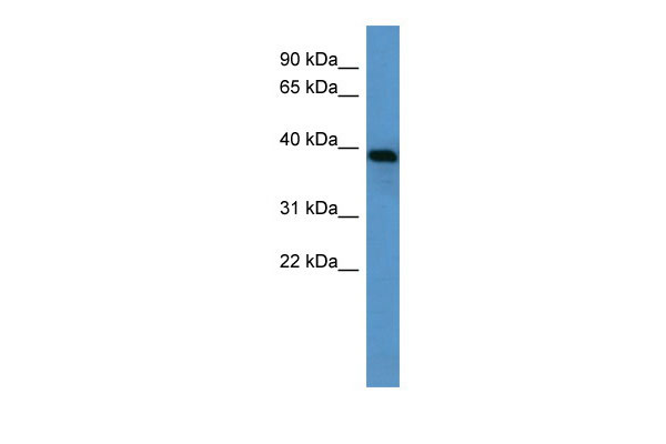
Ambp antibody - N-terminal region
Rabbit Polyclonal Antibody
- 产品详情
- 实验流程
Application
| WB |
|---|---|
| Primary Accession | Q64240 |
| Other Accession | NM_012901, NP_037033 |
| Reactivity | Human, Mouse, Rat, Rabbit, Pig, Dog, Horse, Bovine |
| Predicted | Human, Mouse, Rat, Pig, Bovine |
| Host | Rabbit |
| Clonality | Polyclonal |
| Calculated MW | 38851 Da |
| Gene ID | 25377 |
|---|---|
| Alias Symbol | Ambp |
| Other Names | Protein AMBP, Alpha-1-microglobulin, Inter-alpha-trypsin inhibitor light chain, ITI-LC, Bikunin, HI-30, Trypstatin, Ambp, Itil |
| Format | Liquid. Purified antibody supplied in 1x PBS buffer with 0.09% (w/v) sodium azide and 2% sucrose. |
| Reconstitution & Storage | Add 50 ul of distilled water. Final anti-Ambp antibody concentration is 1 mg/ml in PBS buffer with 2% sucrose. For longer periods of storage, store at 20°C. Avoid repeat freeze-thaw cycles. |
| Precautions | Ambp antibody - N-terminal region is for research use only and not for use in diagnostic or therapeutic procedures. |
| Name | Ambp |
|---|---|
| Synonyms | Itil |
| Function | [Alpha-1-microglobulin]: Antioxidant and tissue repair protein with reductase, heme-binding and radical-scavenging activities. Removes and protects against harmful oxidants and repairs macromolecules in intravascular and extravascular spaces and in intracellular compartments. Intravascularly, plays a regulatory role in red cell homeostasis by preventing heme- and reactive oxygen species- induced cell damage. Binds and degrades free heme to protect fetal and adult red blood cells from hemolysis. Reduces extracellular methemoglobin, a Fe3+ (ferric) form of hemoglobin that cannot bind oxygen, back to the Fe2+ (ferrous) form deoxyhemoglobin, which has oxygen-carrying potential. Upon acute inflammation, inhibits oxidation of low-density lipoprotein particles by MPO and limits vascular damage. Extravascularly, protects from oxidation products formed on extracellular matrix structures and cell membranes. Catalyzes the reduction of carbonyl groups on oxidized collagen fibers and preserves cellular and extracellular matrix ultrastructures. Importantly, counteracts the oxidative damage at blood-placenta interface, preventing leakage of free fetal hemoglobin into the maternal circulation. Intracellularly, has a role in maintaining mitochondrial redox homeostasis. Bound to complex I of the respiratory chain of mitochondria, may scavenge free radicals and preserve mitochondrial ATP synthesis. Protects renal tubule epithelial cells from heme-induced oxidative damage to mitochondria. Reduces cytochrome c from Fe3+ (ferric) to the Fe2+ (ferrous) state through formation of superoxide anion radicals in the presence of ascorbate or NADH/NADPH electron donor cofactors, ascorbate being the preferred cofactor (By similarity). Has a chaperone role in facilitating the correct folding of bikunin in the endoplasmic reticulum compartment (By similarity). |
| Cellular Location | [Alpha-1-microglobulin]: Secreted {ECO:0000250|UniProtKB:P02760}. Endoplasmic reticulum {ECO:0000250|UniProtKB:P02760}. Cytoplasm, cytosol {ECO:0000250|UniProtKB:P02760}. Cell membrane {ECO:0000250|UniProtKB:P02760}; Peripheral membrane protein {ECO:0000250|UniProtKB:P02760}. Nucleus membrane {ECO:0000250|UniProtKB:P02760}; Peripheral membrane protein {ECO:0000250|UniProtKB:P02760}. Mitochondrion inner membrane {ECO:0000250|UniProtKB:P02760}; Peripheral membrane protein {ECO:0000250|UniProtKB:P02760}. Secreted, extracellular space, extracellular matrix {ECO:0000250|UniProtKB:P02760}. Note=The cellular uptake occurs via a non-endocytotic pathway and allows for localization to various membrane structures. A specific binding to plasma membrane suggests the presence of a cell receptor, yet to be identified Directly binds collagen fibers type I. {ECO:0000250|UniProtKB:P02760} |
| Tissue Location | Expressed by the liver and secreted in plasma. |
Research Areas
For Research Use Only. Not For Use In Diagnostic Procedures.
Application Protocols
Provided below are standard protocols that you may find useful for product applications.
终于等到您。ABCEPTA(百远生物)抗体产品。
点击下方“我要评价 ”按钮提交您的反馈信息,您的反馈和评价是我们最宝贵的财富之一,
我们将在1-3个工作日内处理您的反馈信息。
如有疑问,联系:0512-88856768 tech-china@abcepta.com.


