


 癌症的基本特征包括细胞增殖、血管生成、迁移、凋亡逃避机制和细胞永生等。找到癌症发生过程中这些通路的关键标记物和对应的抗体用于检测至关重要。
 为您推荐一个泛素化位点预测神器——泛素化分析工具,可以为您的蛋白的泛素化位点作出预测和评分。
 细胞自噬受体图形绘图工具为你的蛋白的细胞受体结合位点作出预测和评分,识别结合到自噬通路中的蛋白是非常重要的,便于让我们理解自噬在正常生理、病理过程中的作用,如发育、细胞分化、神经退化性疾病、压力条件下、感染和癌症。
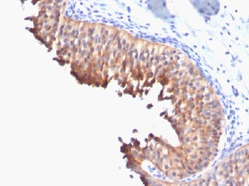
Anti-Cytokeratin 10 Antibody
Mouse Monoclonal Antibody
- 产品详情
- 实验流程
- 背景知识
Application
| IHC-P, IF, FC |
|---|---|
| Primary Accession | P13645 |
| Other Accession | 99936 |
| Reactivity | Human, Dog |
| Host | Mouse |
| Clonality | Monoclonal |
| Isotype | Mouse / IgG1, kappa |
| Clone Names | DE-K10 |
| Calculated MW | 58827 Da |
| Gene ID | 3858 |
|---|---|
| Other Names | BCIE, BIE, EHK, Keratin Type I Cytoskeletal 10, KRT10 |
| Application Note | Flow Cytometry (0.5-1ug/million cells in 0.1ml); Immunofluorescence (0.5-1ug/ml); ,Immunohistology (Formalin-fixed) (0.1-0.2ug/ml for 30 min at RT),(Staining of formalin-fixed tissues requires boiling tissue sections in 10mM citrate buffer, pH 6.0, for 10-20 min followed by cooling at RT for 20 minutes),Optimal dilution for a specific application should be determined. |
| Format | 200ug/ml of Ab purified from Bioreactor Concentrate by Protein A/G. Prepared in 10mM PBS with 0.05% BSA & 0.05% azide. Also available WITHOUT BSA & azide at 1.0mg/ml. |
| Storage | Store at 2 to 8°C.Antibody is stable for 24 months. |
| Precautions | Anti-Cytokeratin 10 Antibody is for research use only and not for use in diagnostic or therapeutic procedures. |
| Name | KRT10 |
|---|---|
| Synonyms | KPP |
| Function | Plays a role in the establishment of the epidermal barrier on plantar skin (By similarity). Involved in the maintenance of cell layer development and keratin filament bundles in suprabasal cells of the epithelium (By similarity). |
| Cellular Location | Secreted, extracellular space. Cell surface. Cytoplasm |
| Tissue Location | Seen in all suprabasal cell layers including stratum corneum. Expressed on the surface of lung cell lines (PubMed:19627498). Localized on the surface of desquamated nasal epithelial cells (at protein level) (PubMed:12427098) |
For Research Use Only. Not For Use In Diagnostic Procedures.
Provided below are standard protocols that you may find useful for product applications.
BACKGROUND
This MAb recognizes a protein of 56.5kDa, identified as cytokeratin 10 (CK10). CK10 is expressed in all suprabasal layers of the epidermis. In the epidermis, expression of CK10 strictly parallels the extent of differentiation; it is absent in the basal layer, appears in the first suprabasal layers and increases in concentration towards the granular layer. However, CK10 is rarely detected in early stages of vulvar squamous carcinomas (tumors less than 2 cm, clinical stage I) regardless of the tumor grade. In larger and more advanced tumors (greater than 2 cm, clinical stages II and III), CK10 is detected very frequently. Expression of CK10 is related to maturation of malignant keratinocytes, being preferentially detected in more-differentiated parts.
终于等到您。ABCEPTA(百远生物)抗体产品。
点击下方“我要评价 ”按钮提交您的反馈信息,您的反馈和评价是我们最宝贵的财富之一,
我们将在1-3个工作日内处理您的反馈信息。
如有疑问,联系:0512-88856768 tech-china@abcepta.com.


