


 癌症的基本特征包括细胞增殖、血管生成、迁移、凋亡逃避机制和细胞永生等。找到癌症发生过程中这些通路的关键标记物和对应的抗体用于检测至关重要。
 为您推荐一个泛素化位点预测神器——泛素化分析工具,可以为您的蛋白的泛素化位点作出预测和评分。
 细胞自噬受体图形绘图工具为你的蛋白的细胞受体结合位点作出预测和评分,识别结合到自噬通路中的蛋白是非常重要的,便于让我们理解自噬在正常生理、病理过程中的作用,如发育、细胞分化、神经退化性疾病、压力条件下、感染和癌症。
TLR6 Antibody (C-term)
Purified Rabbit Polyclonal Antibody (Pab)
- 产品详情
- 文献引用 : 1
- 实验流程
- 背景知识
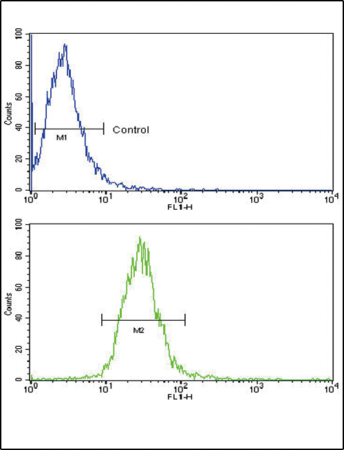
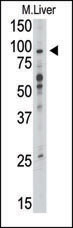
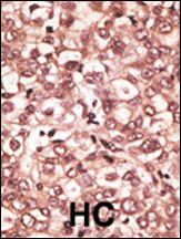
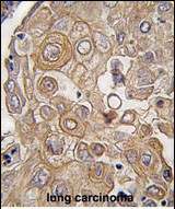
Application
| FC, WB, IHC-P, E |
|---|---|
| Primary Accession | Q9Y2C9 |
| Reactivity | Human |
| Host | Rabbit |
| Clonality | Polyclonal |
| Isotype | Rabbit IgG |
| Calculated MW | 91880 Da |
| Antigen Region | 393-423 aa |
| Gene ID | 10333 |
|---|---|
| Other Names | Toll-like receptor 6, CD286, TLR6 |
| Target/Specificity | This TLR6 antibody is generated from rabbits immunized with a KLH conjugated synthetic peptide between 393-423 amino acids from the C-terminal region of human TLR6. |
| Dilution | FC~~1:10~50 WB~~1:1000 IHC-P~~1:100~500 E~~Use at an assay dependent concentration. |
| Format | Purified polyclonal antibody supplied in PBS with 0.09% (W/V) sodium azide. This antibody is prepared by Saturated Ammonium Sulfate (SAS) precipitation followed by dialysis against PBS. |
| Storage | Maintain refrigerated at 2-8°C for up to 2 weeks. For long term storage store at -20°C in small aliquots to prevent freeze-thaw cycles. |
| Precautions | TLR6 Antibody (C-term) is for research use only and not for use in diagnostic or therapeutic procedures. |
| Name | TLR6 |
|---|---|
| Function | Participates in the innate immune response to Gram-positive bacteria and fungi. Specifically recognizes diacylated and, to a lesser extent, triacylated lipopeptides (PubMed:20037584). In response to diacylated lipopeptides, forms the activation cluster TLR2:TLR6:CD14:CD36, this cluster triggers signaling from the cell surface and subsequently is targeted to the Golgi in a lipid-raft dependent pathway (PubMed:16880211). Acts via MYD88 and TRAF6, leading to NF-kappa-B activation, cytokine secretion and the inflammatory response. Recognizes mycoplasmal macrophage-activating lipopeptide-2kD (MALP-2), soluble tuberculosis factor (STF), phenol-soluble modulin (PSM) and B.burgdorferi outer surface protein A lipoprotein (OspA-L) cooperatively with TLR2 (PubMed:11441107). In complex with TLR4, promotes sterile inflammation in monocytes/macrophages in response to oxidized low-density lipoprotein (oxLDL) or amyloid-beta 42. In this context, the initial signal is provided by oxLDL- or amyloid-beta 42- binding to CD36. This event induces the formation of a heterodimer of TLR4 and TLR6, which is rapidly internalized and triggers inflammatory response, leading to the NF-kappa-B-dependent production of CXCL1, CXCL2 and CCL9 cytokines, via MYD88 signaling pathway, and CCL5 cytokine, via TICAM1 signaling pathway, as well as IL1B secretion (PubMed:11441107, PubMed:20037584). |
| Cellular Location | Cell membrane; Single-pass type I membrane protein. Cytoplasmic vesicle, phagosome membrane {ECO:0000250|UniProtKB:Q9EPW9}; Single-pass type I membrane protein. Membrane raft. Golgi apparatus. Note=Upon complex formation with CD36 and TLR4, internalized through dynamin-dependent endocytosis. Does not reside in lipid rafts before stimulation but accumulates increasingly in the raft upon the presence of the microbial ligand. In response to diacylated lipoproteins, TLR2:TLR6 heterodimers are recruited in lipid rafts, this recruitment determine the intracellular targeting to the Golgi apparatus (PubMed:16880211). |
| Tissue Location | Detected in monocytes, CD11c+ immature dendritic cells, plasmacytoid pre-dendritic cells and dermal microvessel endothelial cells |
For Research Use Only. Not For Use In Diagnostic Procedures.

Provided below are standard protocols that you may find useful for product applications.
BACKGROUND
TLR6, a Type I membrane protein that belongs to the Toll-like rceptor family, participates in the innate immune response to Gram-positive bacteria and fungi. It acts via MyD88 and TRAF6, leading to NF-kappa-B activation, cytokine secretion and the inflammatory response. The protein recognizes mycoplasmal macrophage-activating lipopeptide-2kD (MALP-2), soluble tuberculosis factor (STF), phenol-soluble modulin (PSM) and B.burgdorferi outer surface protein A lipoprotein (OspA-L) cooperatively with TLR2. It binds to TLR2 via their respective extracellular domains, and to MyD88 via their respective TIR domains. TLR6 is detected in monocytes, CD11c+ immature dendritic cells, plasmacytoid pre-dendritic cells and dermal microvessel endothelial cells.
REFERENCES
Bulut, Y., et al., J. Immunol. 167(2):987-994 (2001).
Takeuchi, O., et al., Gene 231 (1-2), 59-65 (1999).
终于等到您。ABCEPTA(百远生物)抗体产品。
点击下方“我要评价 ”按钮提交您的反馈信息,您的反馈和评价是我们最宝贵的财富之一,
我们将在1-3个工作日内处理您的反馈信息。
如有疑问,联系:0512-88856768 tech-china@abcepta.com.


