


 癌症的基本特征包括细胞增殖、血管生成、迁移、凋亡逃避机制和细胞永生等。找到癌症发生过程中这些通路的关键标记物和对应的抗体用于检测至关重要。
 为您推荐一个泛素化位点预测神器——泛素化分析工具,可以为您的蛋白的泛素化位点作出预测和评分。
 细胞自噬受体图形绘图工具为你的蛋白的细胞受体结合位点作出预测和评分,识别结合到自噬通路中的蛋白是非常重要的,便于让我们理解自噬在正常生理、病理过程中的作用,如发育、细胞分化、神经退化性疾病、压力条件下、感染和癌症。
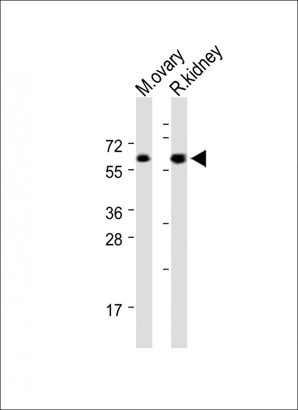
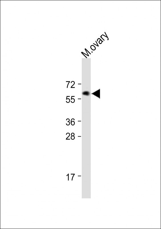
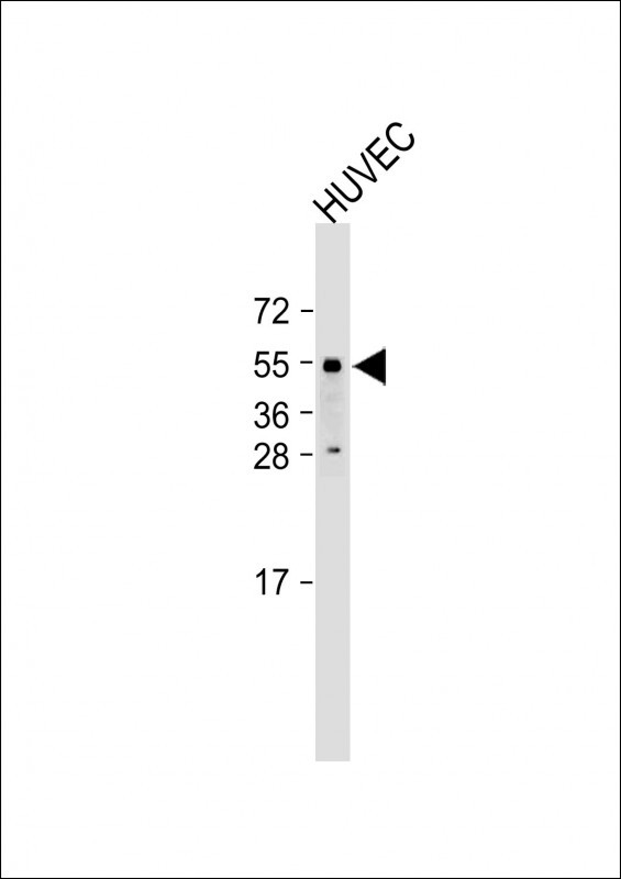
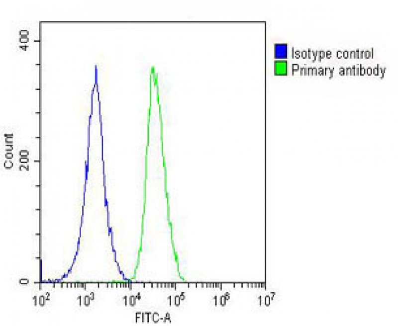
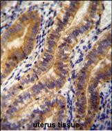
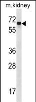
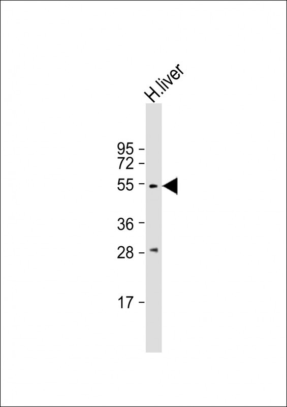
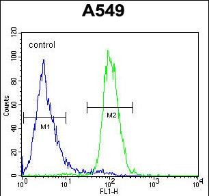
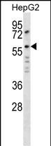
ANGPT2 Antibody (C-term)
Affinity Purified Rabbit Polyclonal Antibody (Pab)
- 产品详情
- 文献引用 : 2
- 实验流程
- 背景知识
Application
| WB, IHC-P, FC, E |
|---|---|
| Primary Accession | O15123 |
| Other Accession | NP_001138.1 |
| Reactivity | Human, Mouse, Rat |
| Host | Rabbit |
| Clonality | Polyclonal |
| Isotype | Rabbit IgG |
| Calculated MW | 56919 Da |
| Antigen Region | 404-432 aa |
| Gene ID | 285 |
|---|---|
| Other Names | Angiopoietin-2, ANG-2, ANGPT2 |
| Target/Specificity | This ANGPT2 antibody is generated from rabbits immunized with a KLH conjugated synthetic peptide between 404-432 amino acids from the C-terminal region of human ANGPT2. |
| Dilution | WB~~1:1000 IHC-P~~1:100~500 FC~~1:10~50 E~~Use at an assay dependent concentration. |
| Format | Purified polyclonal antibody supplied in PBS with 0.05% (V/V) Proclin 300. This antibody is prepared by Saturated Ammonium Sulfate (SAS) precipitation followed by dialysis against PBS. |
| Storage | Maintain refrigerated at 2-8°C for up to 2 weeks. For long term storage store at -20°C in small aliquots to prevent freeze-thaw cycles. |
| Precautions | ANGPT2 Antibody (C-term) is for research use only and not for use in diagnostic or therapeutic procedures. |
| Name | ANGPT2 |
|---|---|
| Function | Binds to TEK/TIE2, competing for the ANGPT1 binding site, and modulating ANGPT1 signaling (PubMed:15284220, PubMed:19116766, PubMed:19223473, PubMed:9204896). Can induce tyrosine phosphorylation of TEK/TIE2 in the absence of ANGPT1 (PubMed:15284220, PubMed:19116766, PubMed:19223473, PubMed:9204896). In the absence of angiogenic inducers, such as VEGF, ANGPT2-mediated loosening of cell-matrix contacts may induce endothelial cell apoptosis with consequent vascular regression. In concert with VEGF, it may facilitate endothelial cell migration and proliferation, thus serving as a permissive angiogenic signal (PubMed:15284220, PubMed:19116766, PubMed:19223473, PubMed:9204896). Involved in the regulation of lymphangiogenesis (PubMed:32908006). |
| Cellular Location | Secreted. |
For Research Use Only. Not For Use In Diagnostic Procedures.

Provided below are standard protocols that you may find useful for product applications.
BACKGROUND
The protein encoded by this gene is an antagonist of angiopoietin 1 (ANGPT1) and endothelial TEK tyrosine kinase (TIE-2, TEK). The encoded protein disrupts the vascular remodeling ability of ANGPT1 and may induce endothelial cell apoptosis. Three transcript variants encoding three different isoforms have been found for this gene.
REFERENCES
Morrissey, C., et al. Prostate 70(16):1799-1808(2010) Romero, R., et al. Am. J. Obstet. Gynecol. 203 (4), 361 (2010) : Bento, C.F., et al. Exp. Physiol. 95(9):955-970(2010) Vrbacky, F., et al. Hematology 15(4):210-214(2010) Chen, J., et al. Biochem. Biophys. Res. Commun. 398(2):212-216(2010)
终于等到您。ABCEPTA(百远生物)抗体产品。
点击下方“我要评价 ”按钮提交您的反馈信息,您的反馈和评价是我们最宝贵的财富之一,
我们将在1-3个工作日内处理您的反馈信息。
如有疑问,联系:0512-88856768 tech-china@abcepta.com.


